- Conditions
- Myositis ossificans
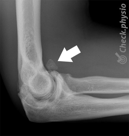
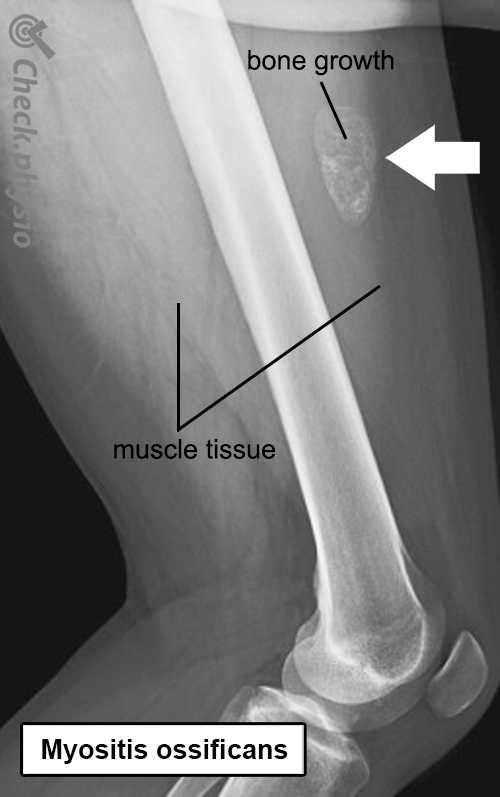
Myositis ossificans Bone growth in a muscle
Introduction
When bone forms in a muscle, this is referred to as myositis ossificans. This is usually the result of muscle contusion or bruising in which a hematoma is converted into bone tissue. A large hematoma after a muscle contusion leads to myositis ossificans in 10 to 20% of cases.
Myositis ossificans may occur in any muscle, but it is most commonly seen in the thigh area after falling on it or perhaps receiving a kick in the thigh. It is therefore relatively more common in people who play contact sports.
Description of condition
Myositis ossificans is a disorder where muscle bruising results in the local formation of bone tissue within that muscle. After having suffered a (severe) contusion, a hematoma develops in the muscle. The body responds to this by triggering an inflammatory response in the muscle (myositis). This can cause warmth, redness, pain and swelling. This inflammatory reaction makes sure that the recovery process starts and the hematoma is eliminated. Most of the time, this happens quite fast and the symptoms disappear in a few weeks. But this does not happen with myositis ossificans. The hematoma calcifies and a small piece of bone forms. This process is referred to as ossification.
In principle, the disorder can develop in anyone and at any age. However, myositis ossificans is seen most in young men between the ages of 20-30 who play contact sports.
Cause and history
Bone usually forms in the muscles after a muscle contusion as a result of contact trauma. This can happen for instance on the soccer field when getting "kneed" in the thigh. However, there is also a rarer cause of myositis ossificans. IT can actually develop in someone who has difficulty moving, for example, due to a coma or paralysis of a body part.
Most of the time, a muscle contusion heals relatively quickly and without problems. However, something different happens with myositis ossificans. The resulting hematoma gradually converts to bone tissue. This process starts about a week after the muscle contusion and finishes six to seven weeks later. The cause of this disorder is unknown and the severity of the symptoms may vary greatly, depending on the location and size of the injury.
Even though the cause is unknown, there are a number of factors that increase the risk of developing myositis ossificans. The likelihood increases with the severity of the contusion and if the mobility of the joint where the muscle runs to, is strongly reduced. Massage and heat immediately after injury can also stimulate bone formation. It is not known why some people do and others do not develop this disorder.
Signs & symptoms
- Hard swelling in the muscle
- Increased pain at night, on/after getting up and during activity.
- Increasingly limited range of movement
- (Visible) hematoma.
Diagnosis
The doctor or physiotherapist will question the patient about the problem. In myositis ossificans, the symptoms usually arise after a muscle contusion that does not start to heal within two weeks. On physical examination, a hard painful lump can be felt in the muscle and the mobility of the joint(s) to which the muscle runs is reduced. At an early stage, an X-ray will not show any abnormalities yet, but the disorder becomes visible after three to four weeks. An ultrasound shows the disorder at a rather earlier stage.
Treatment and recovery
When faced with myositis ossificans, little can be done to promote recovery. It’s a disorder that generally disappears spontaneously. The body needs some time to break down the newly formed superfluous bone. This process starts about six to eight weeks after the muscle contusion.
It progresses slowly and may take months or even up to a year. In other words, treatment consists mostly of rest and patience. A small portion of the bone tissue may remain in the muscle. Luckily, this will not necessarily cause permanent symptoms.
In order to keep joints and muscles of the affected body part flexible, it is advised to do gentle mobility exercises and stay below the pain threshold. Massage, stretching exercises and intensive activities are not recommended.
More info
You can check your symptoms using the online physiotherapy check or make an appointment with a physiotherapy practice in your locality.
References
Nugteren, K. van & Winkel, D. (2012) Onderzoek en behandeling van sportblessures. Onderste extremiteit Houten: Bohn Stafleu van Loghum.
Brukner, P. & Khan, K. (2010) Clinical sports medicine McGraw-Hill: Australia. 3e druk.
Brukner, P. & Khan, K. (2016) Clinical sports medicine (Nederlandse bewerking) 4th edition. Michel van Troost. PreVision, Eindhoven.
Torrance, D. A. & deGraauw, C. (2011) Treatment of post-traumatic myositis ossificans of the anterior thigh with extracorporeal shock wave therapy J Can Chiropr Assoc 2011; 55(4).